Research

Our interdisciplinary MURI team (physics, chemistry, neuroscience, cell biology and dermatology) discovered that Biochemical and Mechanical Systems in cells are also excitable, complementing the faster and larger scale excitable systems character of membrane potentials in neurons. Excitable systems have powerful capabilities, including the ability to exhibit sudden bursts, self-sustaining intermittent or rhythmic dynamics, and collective dynamics organized in space and time. Notably excitable systems can store and transmit information in their dynamics in both space and time across multiple scales.

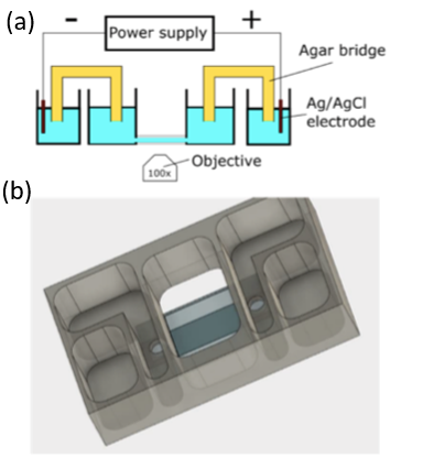
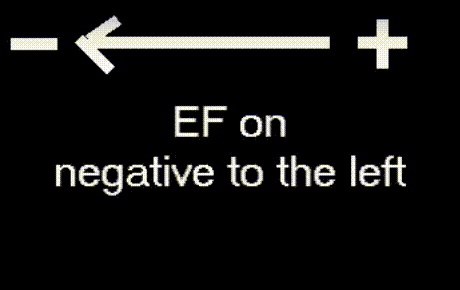
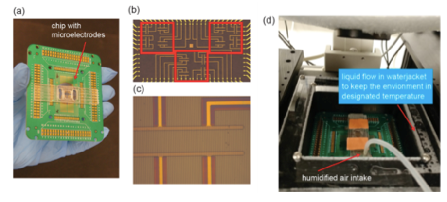
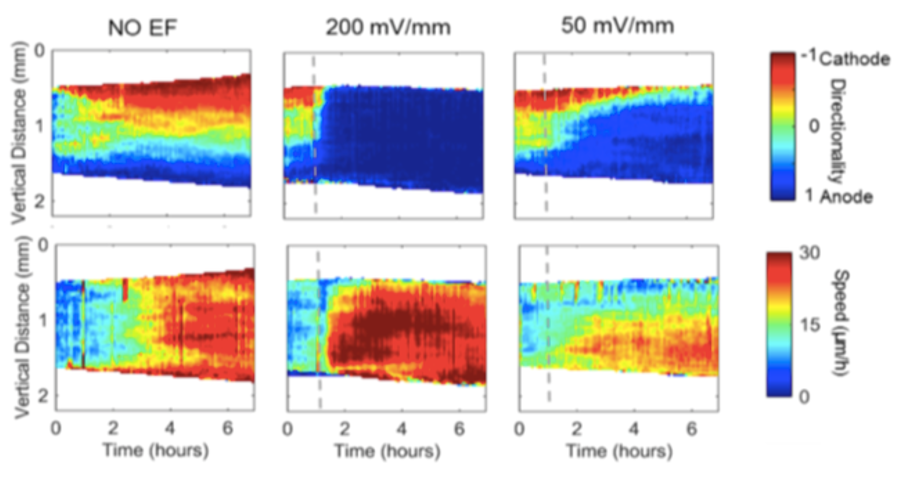
We discovered that the biochemical and biomechanical excitable systems can be precisely modulated and actuated with new synthetic biology switches, through the nanotopography of the environment, and with electric fields (EF). This broad range of actuators exert precise control over living systems, e.g. selective activation of intracellular signaling pathways, cell migration, gene expression, and neuronal activity.


Perturbations with Global Electric Fields


An electrotaxis chamber (left figure) was developed that allows us to study the cells in global electric fields using a broad range of biosensors including PI3K, Ras, and cytoskeleton sensors. Cell migration and excitable waves inside cells are studied under the guidance of global electric field.

Electrotaxis Chamber Design


Cells Migrating in an Electric Field

Left: Electrotaxis chamber design. (a) Schematic of electrotaxis setup for live observation of biosensor responses to an electric field (b) Model that has been used to produce chambers in 3D printer.
Right: A movie taken by Min Zhao's lab shows that cell motion is guided by a global electric field.


Local and Spatio-temporal EF Triggering of Biochemical and Mechanical Cell Behavior


Integrated chip with nanoscale topological pattern aligned with microelectrodes for local mechanical and EF stimulation studies:
We have developed procedures to fabricate customized sub-micron topological features that are aligned with microelectrode arrays on the same glass coverslip substrate so that the response from the cells to simultaneous mechanical and electrical stimuli can be studied in situ .

Integrated chip with nanoscale topological pattern aligned with microelectrodes for local mechanical and EF stimulation studies

Chip and assembled chamber with topological pattern and microelectrode arrays. (a) Chip assembly. (b) Microelectrode pairs with nanoscale ridges. (c) Nanoridges are 0.7 µm wide with a 2 µm gap and are aligned with the electrode. (d) On-stage incubation system for in situ and long term imaging experiments (top cap and wires removed).


Stimulation of Collective Cell Behavior


When a constraint is removed, confluent cells migrate directionally into the available space. How migration directionality and speed increases are initiated at the leading edge and propagate into neighboring cells is not well understood. Using a quantitative technique—Particle Image Velocimetry (PIV)—we revealed that migration directionality and speed had strikingly different dynamics.

PIV analysis of cells migrating in an electric field

Directionality and speed waves propagate in migrating cell sheets. Color codes indicating directionality and speed are shown on the right. When EF lines are parallel with the free edge (perpendicular with the original directional migration), cells at the edge migrate mainly in the direction that follows the principle of vector superposition (vector accumulation/vector addition). The electric field interacts with the motility wave that emerges in the process of cell sheet expansion and disturbs the motility wave both spatially and temporally.